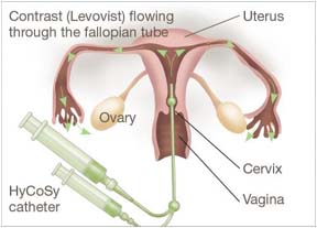
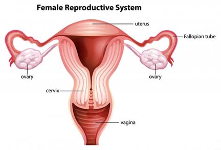
Hysterosalpingo-contrast-sonography (usually shortened toHyCoSy) is a simple and well-tolerated outpatient ultrasound procedure used to assess the patency of the fallopian tubes, as well as detect abnormalities of the uterus and endometrium.
gkbLVjkslkfYiaxks&daVªkLV&lksuksxzkQh ¼vkerkSj ij gkbdkslh ls NksVk½ ,d ljy vkSj vPNh rjg ls lgu'khy vkmV is'ksaV vYVªklkmaM çfØ;k gS tks Qyksfi;u Vîwcksa dh isVsalh dk vkdyu djus ds fy, mi;ksx dh tkrh gS] lkFk gh xHkkZ'k; vkSj ,aMksesfVª;e dh vlkekU;rkvksa dk irk yxkrh gSA
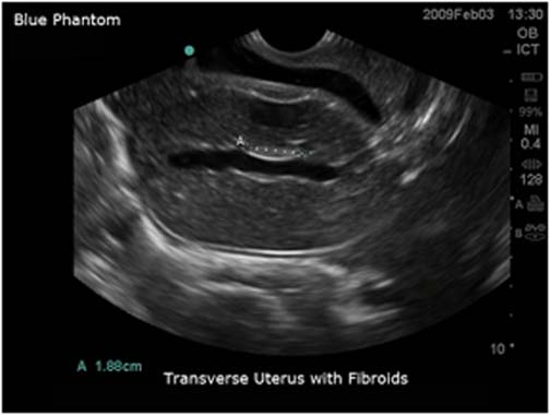
Hydro hysterosonography:
Hysterosonography, which is also called sonohysterography, is a new noninvasive technique that involves the slow infusion of sterile saline solution into a woman's uterus during ultrasound imaging. Hysterosonography allows the doctor to evaluate abnormal growths inside the uterus; abnormalities of the tissue lining the uterus (the endometrium); or disorders affecting deeper tissue layers. Hysterosonography does not require either radiation or contrast media, or invasive surgical procedures
gkbLVjkslksuksxzkQh] ftls lksuksbLVsjksxzkQh Hkh dgk tkrk gS] ,d ubZ noninvasive rduhd gS ftlesa vYVªklkmaM besftax ds nkSjku ,d efgyk ds xHkkZ'k; esa ck¡> uedhu lek/kku ds /khes tylsd 'kkfey gSA gkbLVjkslksuksxzkQh M‚DVj dks xHkkZ'k; ds vanj vlkekU; o`f) dk ewY;kadu djus dh vuqefr nsrh gS; xHkkZ'k; ¼,aMksesfVª;e½ vLrj Ård dh vlkekU;rkvksa; ;k xgjs Ård ijrksa dks çHkkfor djus okys fodkjA Hysterosonography ;k rks fofdj.k ;k foijhr ehfM;k] ;k vkØked 'kY; fpfdRlk çfØ;kvksa dh vko';drk ugha gSA
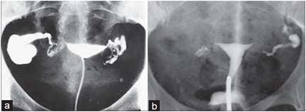
Sion test:
Sonosalpingography (SSG), also known as Sion test, is a diagnostic procedure primarily used for evaluating patency of fallopian tubes. It was introduced as a screening procedure for infertility investigations. It is becoming more popular among practitioners due to absence of side effects.
lksukslkfYiaxksxzkQh ¼,l,lth½] ftls fl;ksu VsLV Hkh dgk tkrk gS] eq[; :i ls QSyksfi;u Vîwcksa dh isVsalh dk ewY;kadu djus ds fy, mi;ksx dh tkus okyh uSnkfud çfØ;k gSA bls cka>iu tkap ds fy, ,d LØhfuax çfØ;k ds :i esa is'k fd;k x;k FkkA nq"çHkkoksa dh vuqifLFkfr ds dkj.k ;g fpfdRldksa ds chp vf/kd yksdfç; gks jgk gSA
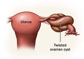
Ovarian torsion:
Ovarian torsion is an uncommon cause of acute abdominal pain in females, but it is a gynecologic emergency - diagnostic delay can result in loss of the ovary. The ovary, and often the fallopian tube as well (adnexal torsion) become twisted around their vascular pedicle.
efgykvksa ds vksosfj;u esa rhoz isV nnZ ,d vlkekU; dkj.k gS] ysfdu ;g ,d L=h jksx laca/kh vkikrdkyhu & Mk;XuksfLVd nsjh ds ifj.kkeLo:i vaMk'k; dk uqdlku gks ldrk gSA vaMk'k;] vkSj vDlj QSyksfi;u Vîwc Hkh ¼,MusDly Vksjlu½ muds laoguh isfMdy ds pkjksa vksj ew¡M+ tkrh gSA
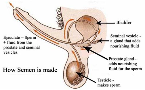
Ejaculation:
Ejaculation is the discharge of semen (usually carrying sperm) from the male reproductory tract. It usually occurs during an orgasm. It is usually the final stage and natural objective of male sexual stimulation, and an essential component of natural conception. In rare cases, ejaculation occurs because of prostatic disease. Ejaculation may also occur spontaneously during sleep (a nocturnal emission or "wet dream").
esy fjçksMfDVo VªSDV ls LieZ ¼vkerkSj ij 'kqØk.kq ys tkus½ dk fuoZgu gksrk gSA ;g vkerkSj ij ,d laHkksx ds nkSjku gksrk gSA ;g iq#"k ;kSu mÙkstuk dk vafre pj.k vkSj çk—frd vo/kkj.kk dk ,d vko';d ?kVd gSA nqyZHk ekeyksa esa] çksLVsfVd chekjh ds dkj.k Hkh uhan ds nkSjku L[kyu gksrk gSA
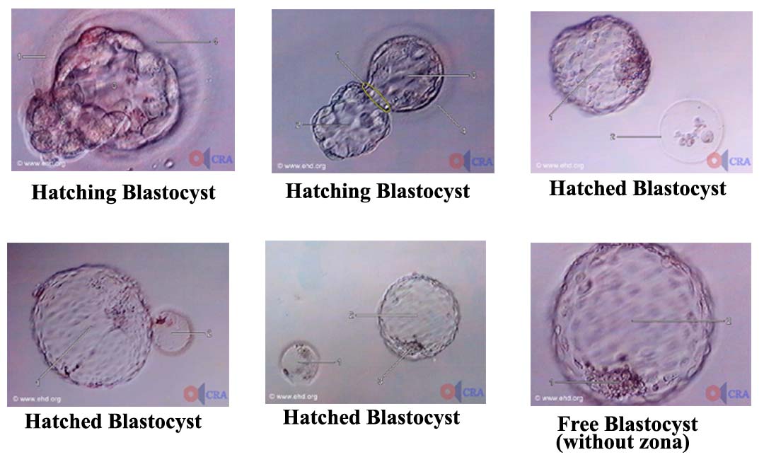
Blastocyst Hatching:
Hatching is a phenomenon seen with embryos developing outside the body. Under natural conditions inside the body, the zona is believed to degenerate and disappear after the embryo reaches the uterine cavity and prepares to implant.
gSfpax ,d ?kVuk gS tks 'kjhj ds ckgj fodflr H:.k ds lkFk ns[kh tkrh gSA 'kjhj ds vanj çk—frd ifjfLFkfr;ksa esa] H:.k xHkkZ'k; xqgk rd igqapus ds ckn vkSj çR;kjksi.k ds fy, rS;kj gksus ds ckn tksu dksdegenerate and disappear ekuk tkrk gSA
Label Key:
1. zona pellucida 2. embryo escaping from the zona (called hatching) 3. blastocyst cavity 4. perivitelline space
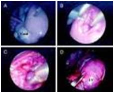
Fetoscopy:
Fetoscopy is an endoscopic procedure during pregnancy to allow access to the fetus, the amniotic cavity, the umbilical cord, and the fetal side of the placenta. A small (3–4 mm) incision is made in the abdomen, and an endoscope is inserted through the abdominal wall and uterus into the amniotic cavity.
Fetoscopy is a procedure that is carried out on pregnant women with the help of a fetoscope. A fetoscope is a thin and flexible medical apparatus. It carries out an endoscopic pregnancy test. The fetoscope is inserted into the uterus through an incision made in the stomach. An ultra sound is required when carrying out a fetoscopy. It ensures that the doctor can see where the incision is being carried out to avoid harming the baby and also how the fetoscope is moving in the womb. Fetoscopy is used to judge progress of the pregnancy or treat any abnormalities. The Amniocentesis procedure is another procedure that offers information during the prenatal period of the status of the fetus.